Page: 1
/ 14
Total 165 questions
ARDMS Abdomen Sonography Examination AB-Abdomen Exam Questions
Question 1
Which anatomical area of the male reproductive system is demonstrated in this endorectal image?
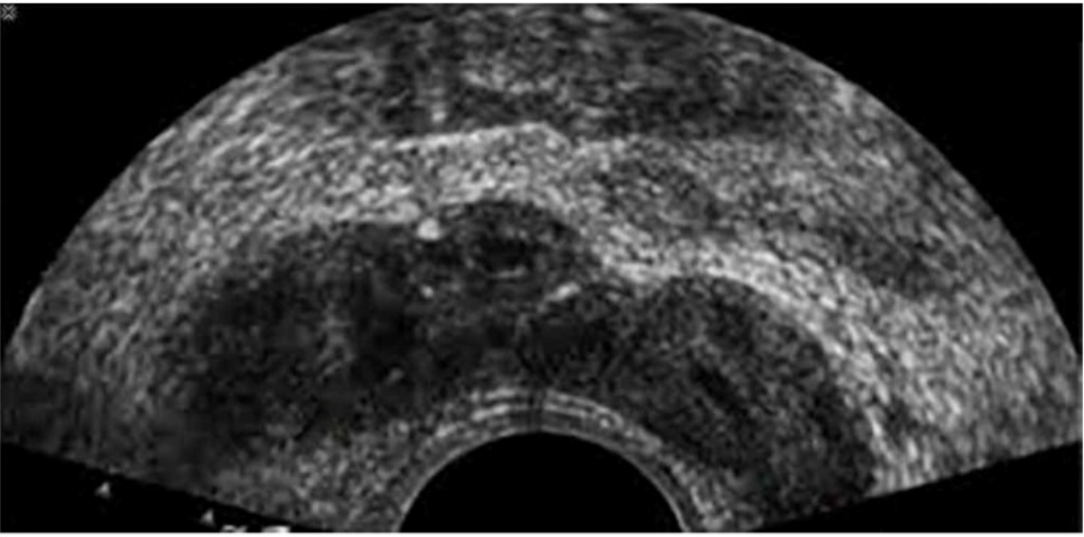
Answer : C
The ultrasound image shown is a transverse endorectal (transrectal) ultrasound, commonly used to evaluate the prostate and adjacent structures. The two hypoechoic (dark) oval-shaped structures seen superior and posterior to the prostate are characteristic of the seminal vesicles.
The seminal vesicles are paired, elongated glands located superior and posterior to the base of the prostate and are best visualized in transverse planes on endorectal imaging. They appear as hypoechoic or anechoic structures with internal septations, depending on the degree of fluid content.
In contrast:
The urethra appears as a central echogenic linear structure within the prostate.
The prostate base is more inferior in the scan and is visualized just above the urethra.
The ejaculatory ducts are usually not as prominently visualized and are located medial to the seminal vesicles, entering the prostate near the verumontanum.
This image most clearly demonstrates the bilateral seminal vesicles.
Rumack CM, Wilson SR, Charboneau JW, Levine D. Diagnostic Ultrasound, 5th ed. Elsevier; 2017.
ACR--AIUM--SRU Practice Parameter for the Performance of an Ultrasound Examination of the Prostate (2021).
Hagen-Ansert SL. Textbook of Diagnostic Sonography, 8th ed. Elsevier; 2017.
Question 2
What is the main purpose for performing focused abdominal sonography for trauma (FAST) exams?
Answer : A
The FAST exam is primarily used to detect free intraperitoneal or pericardial fluid in trauma patients, serving as a rapid, bedside assessment tool. While organ injuries may be suspected, the FAST exam is not primarily designed to assess for solid organ lacerations.
According to AIUM and ACEP guidelines:
''The primary goal of the FAST exam is to detect the presence of free fluid suggestive of hemorrhage in trauma patients.''
American College of Emergency Physicians (ACEP) Ultrasound Guidelines, 2016.
AIUM Practice Parameter for the Performance of the FAST Examination, 2020.
---
Question 3
Which abnormality is the most common adult adrenal tumor?
Answer : D
Adrenal adenomas are the most common adrenal tumors in adults. They are often discovered incidentally (adrenal incidentalomas) and are usually nonfunctioning, though some may secrete cortisol or aldosterone. Neuroblastoma is common in children, pheochromocytomas are rarer catecholamine-producing tumors, and adrenal cortical carcinoma is malignant but much less common than adenomas.
According to Rumack's Diagnostic Ultrasound:
''Adrenal adenomas are the most common adrenal masses in adults, frequently identified incidentally on imaging studies.''
Rumack CM, Wilson SR, Charboneau JW, Levine D. Diagnostic Ultrasound. 5th ed. Elsevier, 2017.
ACR Incidental Findings Committee Guidelines for Adrenal Masses, 2017.
---
Question 4
Which sonographic feature is typical of a thyroid adenoma?
Answer : B
Thyroid adenomas typically present as well-defined nodules surrounded by a thin, hypoechoic peripheral halo representing compressed thyroid parenchyma or fibrous capsule. Irregular margins suggest malignancy.
According to Rumack's Diagnostic Ultrasound:
''A thin hypoechoic halo is characteristic of benign thyroid adenomas.''
Rumack CM, Wilson SR, Charboneau JW, Levine D. Diagnostic Ultrasound. 5th ed. Elsevier, 2017.
AIUM Practice Parameter for Thyroid Ultrasound, 2020.
---
Question 5
Which finding is an indication for renal biopsy to assess for renal failure?
Answer : A
Significant proteinuria, especially if persistent or in the nephrotic range, may indicate glomerular disease and is a common indication for renal biopsy. Leukocytosis and hypercalcemia are not specific for renal biopsy. Hematuria may warrant biopsy if accompanied by proteinuria.
According to KDIGO Clinical Practice Guidelines:
''Persistent proteinuria is one of the strongest indications for renal biopsy to evaluate underlying glomerular pathology.''
Kidney Disease: Improving Global Outcomes (KDIGO) Clinical Practice Guideline for Glomerulonephritis, 2021.
American Society of Nephrology (ASN) Nephrology Board Review, 2021.
Question 6
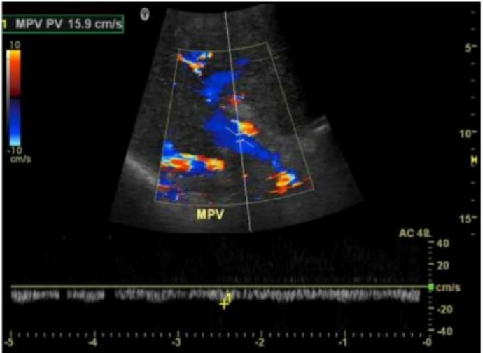
Which condition is demonstrated in this image?
Answer : A
The image shows a color Doppler ultrasound of the main portal vein (MPV), which appears irregular and replaced by multiple small, serpiginous vascular channels --- a hallmark of cavernous transformation. Cavernous transformation of the portal vein is a late complication of chronic portal vein thrombosis, in which collateral vessels develop around the thrombosed portal vein to bypass the obstruction.
Key Doppler ultrasound features of cavernous transformation:
Absence of a normal portal vein
Multiple tortuous vessels in the porta hepatis
Color Doppler shows hepatopetal flow in these channels
Low velocity, continuous waveform flow in collateral vessels
Differentiation from other options:
B . Portal vein thrombosis: Would show an absence of color flow within the portal vein lumen and possibly echogenic material within the vessel. There would be no serpiginous collateral vessels yet if it's an acute process.
C . Portal hypertension: Often diagnosed with other sonographic findings (e.g., splenomegaly, reversed portal flow, varices) but not characterized by the replacement of the portal vein by collateral vessels.
Rumack CM, Wilson SR, Charboneau JW, Levine D. Diagnostic Ultrasound. 5th Edition. Elsevier, 2018. Chapter: Portal Venous System, pp. 107--110.
American Institute of Ultrasound in Medicine (AIUM). Practice Parameter for the Performance of a Vascular Ultrasound Examination, 2021.
Radiopaedia.org. Cavernous transformation of the portal vein: https://radiopaedia.org/articles/cavernous-transformation-of-the-portal-vein
Question 7
In which position should a patient be placed when internal echoes are seen within a fluid-filled bladder?
Answer : C
Lateral decubitus positioning allows shifting of internal echoes within the bladder, helping differentiate mobile debris (such as blood clots or sediment) from adherent masses like tumors. This technique is helpful in evaluating questionable bladder filling defects.
According to Rumack's Diagnostic Ultrasound:
''Changing the patient's position, such as turning to the lateral decubitus, can help distinguish mobile debris from attached bladder wall lesions.''
Rumack CM, Wilson SR, Charboneau JW, Levine D. Diagnostic Ultrasound. 5th ed. Elsevier, 2017.
AIUM Practice Parameter for Bladder Ultrasound, 2020.