Page: 1
/ 14
Total 215 questions
ARDMS Sonography Principles and Instrumentation SPI Exam Questions
Question 1
Which factor does a string phantom evaluate?
Answer : C
Comprehensive and Detailed Explanation From Exact Extract:
The string phantom is designed to evaluate Doppler system performance, particularly flow velocity accuracy. It uses a moving string to simulate constant flow velocities.
Principles and Instrumentation state:
'The string phantom simulates known flow velocities and is used to assess Doppler accuracy in measuring flow velocities.'
Two-dimensional resolution (A) is assessed by test patterns.
Intensity values (B) are tested with hydrophones.
Slice thickness (D) is tested with special slice thickness phantoms.
Therefore, the correct answer is C: Flow velocity.
---
Question 2
Which action may reduce the number of lines in a frame without a loss of temporal resolution?
Answer : B
Narrowing the field of view reduces the number of scan lines that need to be processed per frame. This allows the ultrasound system to maintain or even increase the frame rate without compromising temporal resolution. Temporal resolution, which refers to the system's ability to depict motion accurately, is directly related to the frame rate. Reducing the field of view ensures fewer lines are needed to create each image, thus preserving the frame rate and temporal resolution.
ARDMS Sonography Principles and Instrumentation guidelines
Kremkau, F. W. (2015). Diagnostic Ultrasound: Principles and Instruments.
Question 3
Which resolution is improved by focusing?
Answer : A
Focusing improves lateral resolution in ultrasound imaging. Lateral resolution refers to the system's ability to distinguish between two points that are side by side (perpendicular to the sound beam's path). By focusing the ultrasound beam, the width of the beam is narrowed at the focal point, enhancing the system's ability to resolve structures that are close together in the lateral plane. This results in clearer, more detailed images of the anatomical structures. Reference:
American Registry for Diagnostic Medical Sonography (ARDMS) Sonography Principles and Instrumentation study materials.
Diagnostic Ultrasound: Principles and Instruments by Kremkau, F. W. (latest edition).
Question 4
What is ensemble length (packet size)?
Answer : B
Comprehensive and Detailed Explanation From Exact Extract:
Ensemble length, also known as packet size, refers to the number of ultrasound pulses sent along each scan line when performing color Doppler or power Doppler imaging. Increasing the ensemble length allows for more accurate velocity estimation and improved sensitivity to low flow but comes at the expense of decreased frame rate.
Official sonography instrumentation reference states:
''Ensemble length refers to the number of pulses per scan line used in color Doppler imaging. A larger ensemble improves sensitivity to slow flow and provides more accurate frequency (velocity) estimates but reduces temporal resolution.''
Thus, the correct answer is B: Number of pulses along a scan line.
---
Question 5
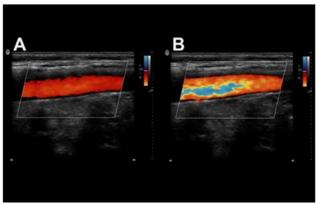
Which color control adjustment caused the change from image A to image B?
Answer : B
Comprehensive and Detailed Explanation From Exact Extract:
In image A, the vessel shows uniform color filling without aliasing. In image B, multiple color bands are present, indicating color aliasing --- where the flow velocities exceeded the Nyquist limit due to a lower velocity scale (PRF).
According to sonography Principles and Instrumentation:
'Aliasing in color Doppler appears when the Doppler shift exceeds the Nyquist limit. Reducing the velocity scale increases the likelihood of aliasing.'
Changing the map (A) would modify the color hues but not introduce aliasing.
Wall filter (C) affects low-velocity signals but not aliasing.
Color packet size (D) affects frame rate and sensitivity, not aliasing directly.
Therefore, the correct answer is B: Velocity scale.
---
Question 6
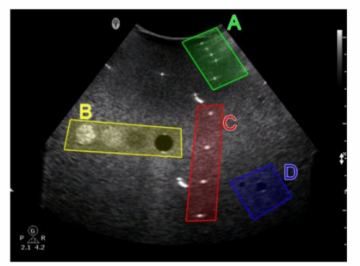
Which target group in this image of a tissue-mimicking phantom is used for gray-scale evaluation?
Answer : C
Gray-scale evaluation in a tissue-mimicking phantom involves assessing the uniformity and accuracy of the gray-scale representation of the tissues.
Option C typically contains structures designed to test the machine's ability to accurately depict varying levels of echogenicity, which is essential for proper gray-scale evaluation.
This area will have a range of echo intensities that help in determining the contrast resolution and the ability of the system to distinguish between different tissue types based on their gray-scale values. Reference:
ARDMS Sonography Principles and Instrumentation guidelines on tissue-mimicking phantoms and image quality evaluation.
Question 7
Which effect does spatial compounding have on ultrasound images?
Answer : D
Comprehensive and Detailed Explanation From Exact Extract:
Spatial compounding acquires multiple frames from different angles and combines them into a single image. This technique reduces the appearance of artifacts such as shadowing and speckle noise, resulting in a smoother, more uniform image.
According to sonography instrumentation reference:
''Spatial compounding reduces artifacts like posterior shadowing and speckle by averaging data from multiple insonation angles.''
Therefore, the correct answer is D: Decreases shadowing.
---
 All Official Question Types